41 art labeling activity structure of a skeletal muscle fiber
Bsc2085l chapter 013 activity 1 skeletal muscle - Course Hero BSC2085L Chapter 013 Activity 1 Skeletal Muscle Organization-005 Part A The area of a sarcomere where the thin actin filaments connect to one another is called the _____. ANSWER: Correct The Z line or Z disc consists of proteins called actinin that anchor the actin filaments together. A message from your instructor... Activity 2: The Neuromuscular Junction Art-labeling Activity: Skeletal ... PDF In this chapter, you will learn that - Pearson 9.2 A skeletal muscle is made up of muscle fibers, nerves, blood vessels, and connective tissues Learning Objective Describe the gross structure of a skeletal muscle. For easy reference, Table 9.1 on p. 286 summarizes the levels of skeletal muscle organi-zation, gross to microscopic, that we describe in this and the following modules.
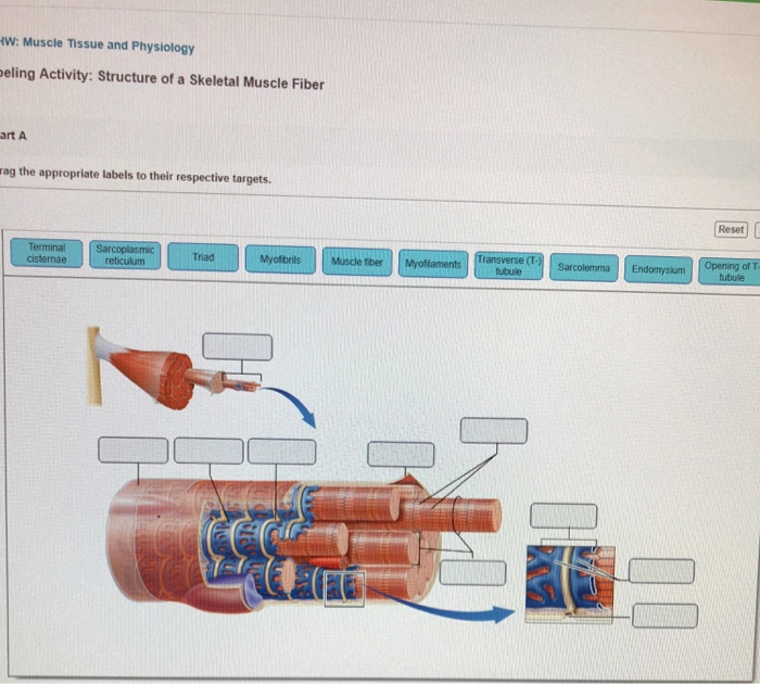
Edit View History Bookmarks Window He... - Anatomy and Physiology Art-Labeling Activity: Structure of a Skeletal Muscle Fiber Part A Drag the appropriate labels to their respective targets. Reset Help Terminal cisternae Myofilaments Transverse (T tubule Triad Endomysium Myofibrils Sarcolemma Sarcoplasmic Opening of T. reticulum tubule Muscle fiber H I Submit Request Answer Provide Feedback OCT . ty a 4 24 Q 80 F3

Art labeling activity structure of a skeletal muscle fiber
Art-labeling activity - structure of skeletal muscle - Chegg This problem has been solved! See the answer. See the answer See the answer done loading. Art-labeling activity: structure of skeletal muscle fiber. Drag the appropriate lablels to their respective targets. Expert Answer. art-labeling activity: the structure of the digestive tract An unregistered player played the game 29 seconds ago. 2018-7-14 Art-labeling Activities Use the art-labeling activities to quiz yourself on key anatomical structures in this chapter. Structural organization of skeletal muscle Reset Help Epimysium Muscle fascicle Endomysium Perimysium Nerve Muscle fibers Blood vessels Tendon Muscle fiber cell. Answered: Art-labeling Activity: Structural… | bartleby match to correct box. Transcribed Image Text: Art-labeling Activity: Structural organization of skeletal muscle Reset Help Epimysium Muscle fascicle Endomysium Perimysium Nerve Muscle fibers Blood vessels Tendon Muscle fiber (cell)
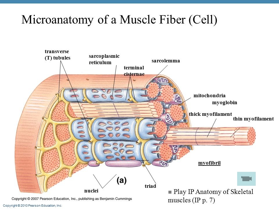
Art labeling activity structure of a skeletal muscle fiber. Answer correct art based question chapter 4 question - Course Hero ANSWER: Correctmultinucleate cells branched cells intercalated discs situated between cells striations tendons and ligaments attached to bones heart ducts of certain glands dense irregular connective tissue smooth muscle tissue skeletal muscle tissue cardiac muscle tissue Bio 2331 Prelab 6 Muscles Part 1.pdf - 2/10/22, 10:55 PM... Art-labeling Activity: The Structure of Skeletal and Cardiac Muscle Fibers Part A Drag the labels to the appropriate location in the figure. ANSWER: Lab Manual Exercise 5 From the Book Pre-lab Quiz Question 9 Part A Specialized cell junctions, called intercalated discs, ... Art-labeling Activity: The Structure of a Skeletal Muscle Fiber Start studying Art-labeling Activity: The Structure of a Skeletal Muscle Fiber. Learn vocabulary, terms, and more with flashcards, games, and other study tools. Search. Create. ... The Structure of a Skeletal Muscle Fiber... OTHER SETS BY THIS CREATOR. Pathophysiology. 11 terms. BabeRuthless0504. Lympathetic System. 37 terms. Skeletal Muscle Fiber Structure and Function - Open Textbooks for Hong Kong Figure 16.18 A skeletal muscle fiber is surrounded by a plasma membrane called the sarcolemma, with a cytoplasm called the sarcoplasm. A muscle fiber is composed of many fibrils packaged into orderly units. The orderly arrangement of the proteins in each unit, shown as red and blue lines, gives the cell its striated appearance.
BIOL.docx - Ch9 Hmwk Art-labeling Activity: Structural ... - Course Hero Art-labeling Activity: Structural organization of skeletal muscle Part A Drag the labels to the appropriate location in the figure. Epimysium Muscle fascicle Endomysium Perimysium Nerve Muscle fibers Blood vessels Tendon Muscle fiber (cell) Help Reset Submit My Answers Give Up Correct Provide FeedbackContinue Art-Labeling Activity: Sarcomere Structure - ARTDCA June 16, 2022 Art-Labeling Activity: Sarcomere Structure. Skeletal muscle matching back of the body. Sarcomere h zone thin (actin) filament thick (myosin) filament z disc z disc m line (c) small part of one myofibril enlarged to show the myofilaments responsible for the banding pattern. PDF The Muscular System Tour Lab The Muscular System - lcboe.net is broken down to provide energy. To help delay muscle fatigue, the muscle fibers are constantly switching on an off to allow individual fibers a moment to rest. This activity will demonstrate the effects of action of muscle fibers. Do this: 1. Hold a popsicle stick in front of you , parallel to the table top. 2. Place a bent paper clip on the ... 10.2 Skeletal Muscle - Anatomy and Physiology 2e | OpenStax These tissues include the skeletal muscle fibers, blood vessels, nerve fibers, and connective tissue. Each skeletal muscle has three layers of connective tissue (called "mysia") that enclose it and provide structure to the muscle as a whole, and also compartmentalize the muscle fibers within the muscle ( Figure 10.3 ).
Solved Art-labeling Activity: The Structure of Skeletal and - Chegg See the answer Art-labeling Activity: The Structure of Skeletal and Cardiac Muscle Fibers Drag the labels to the appropriate location in the figure. Show transcribed image text Expert Answer 100% (2 ratings) The first picture is of a skeletal muscle which can identified by parallel bun … View the full answer 10.2 Skeletal Muscle - Anatomy & Physiology The striations of skeletal muscle are created by the organization of actin and myosin filaments resulting in the banding pattern of myofibrils. These actin and myosin filaments slide over each other to cause shortening of sarcomeres and the cells to produce force. Critical Thinking Questions 1. art-labeling activity: the spinal meninges and associated structures The Structure of a Skeletal Muscle Fiber 2 Terms. The spinal meninges continue upward into the skull where they are continuous with the cranial meninges. The cell body of these neurons are found within the. ... Start studying Art-Labeling Activity. Fill out and upload to BB when done-Mastering Lab Activity Chapter 7-Videos. We would like to ... Muscle Tissue and Physiology Art-Labeling | Chegg.com Question:
Illustration of structure skeletal. Vector illustration of structure ...
chapter 9 Flashcards | Quizlet Art-labeling Activity: The structure of a skeletal muscle fiber PICTURE Chapter Test - Chapter 9 Question 3 Which thin-filament-associated structure is distinguished by its constituents of three globular subunits, one of which has a receptor that binds two calcium ions? a) G-actin b) nebulin c) tropomyosin d) troponin D ...
Anatomy And Physiology Archive | March 14, 2021 | Chegg.com
Week 3 Chapter 9.pdf - 4/23/22, 5:03 PM Week 3 Chapter 9... The tension produced by a contracting skeletal muscle fiber results from the interaction between the thick and thin filaments within sarcomeres. The mechanism of skeletal muscle contraction is explained by the sliding filament theory Read through Spotlight Figure 9.7, and then complete the questions and activity below. Part A - Initiation of Contraction Contraction is initiated by release of ...

Skeletal Muscle Photos and Premium High Res Pictures - Getty Images
Muscle Fibers Teaching Resources | Teachers Pay Teachers Muscle Types: Smooth and Skeletal Muscle Fibers. by. Engaging Science Labs. $3.00. PDF. Study both smooth and skeletal muscles by looking at raw and cooked meat. You should be able to see the fascia and how skeletal muscle is comprised of bundled fibers whereas smooth muscle is not.Questions Answered:What are some characteristics of skeletal ...
Muscles - Runyan-Grigsby's Science Page
A&P 1- CHAPTER 9 MASTERING ASSIGNMENTS Flashcards - Quizlet Art-labeling Activity: The structure of a skeletal muscle fiber PICTURE Which thin filament-associated protein binds two calcium ions? troponin Action potential propagation in a skeletal muscle fiber ceases when acetylcholine is removed from the synaptic cleft.
Solved: Art-labeling Activity: Structure Of Skeletal Muscl... | Chegg.com
Art-Labeling Activity: The Structure Of A Sarcomere Part A Drag The ... Art-Labeling Activity: The Structure Of A Sarcomere Part A Drag The Labels To The Appropriate Location In The Figure. Reset Help A Band Barmere Hand Band MI Art-Labeling Activity: The Structure Of A Skeletal Muscle Fiber Part A Drag The Labels Onto The Diagram To Identity Structural Features Associated With A Skeletal Muscle Fiber.
Post a Comment for "41 art labeling activity structure of a skeletal muscle fiber"