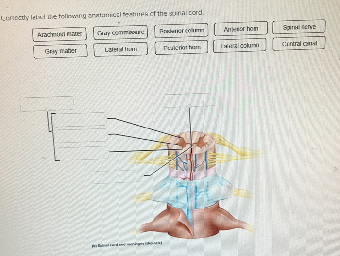
42 correctly label the following anatomical features of the spinal cord.
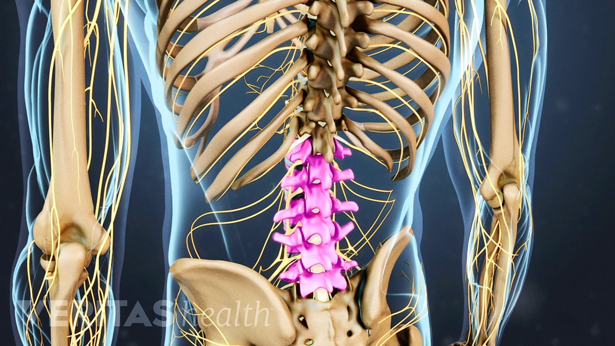
Vertebral Anatomy: Cervical, Thoracic, Lumbar, Sacral Spine The function of the vertebral column, also known as the spinal column, is to the protect and support the spinal cord and bear the weight of the rest of the body. Most individuals are born with a total of 33 vertebrae. They include 7 cervical, 12 thoracic, 5 lumbar, 5 sacral, and 4 coccygeal vertebrae. By adulthood, the 5 sacral vertebrae fuse ... Answered: Anatomy of the Spinal Cord 1. Complete… | bartleby Anatomy of the Spinal Cord 1. Complete the following statements by inserting the proper anatomical terms in the answer blanks. The superior boundary of the spinal cord is at the level of the foramen magnum of the skull, and its inferior boundary is at the level of vertebra.
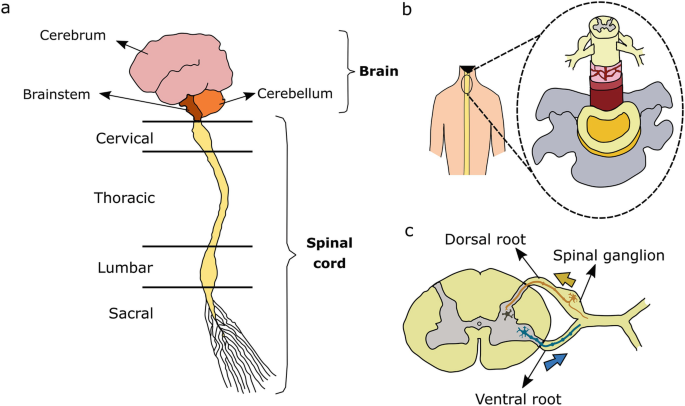
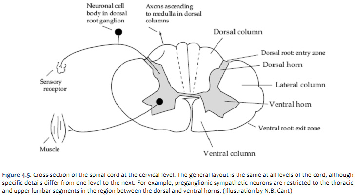
Anatomy of the Spinal Cord (Section 2, Chapter 3) Neuroscience Online ... A dermatome is an area of skin supplied by peripheral nerve fibers originating from a single dorsal root ganglion. If a nerve is cut, one loses sensation from that dermatome. Because each segment of the cord innervates a different region of the body, dermatomes can be precisely mapped on the body surface, and loss of sensation in a dermatome can indicate the exact level of spinal cord damage ...
Correctly label the following anatomical features of the spinal cord.
The deep crease on the anterior surface of the spinal cord is the (a ... Correctly Label The Following Anatomical Features Of The Spinal Cord. Lateral Funiculus Posterior Root Of Spinal Nerve Posterior Funiculus Posterior Horn Anterior Median Fissure Spinal Nerve Gray Commissure Spinal Nerve (B) Spinal Cord And... Correctly Label the Anatomical Features of a Neuron Anatomy of the brain and spinal cord are the most obvious parts of the brain. The nucleus of a neuron is a large, round organ that contains masses of chromatin. The nucleus is also the cell's main organ, and contains the axon and deoxyribonucleic acid (DNA). The axon is the source of the signal in the neuron. Spinal Anatomy | Vertebral Column - SpineUniverse The Atlas is the first cervical vertebra and therefore abbreviated C1. This vertebra supports the skull. Its appearance is different from the other spinal vertebrae. The atlas is a ring of bone made up of two lateral masses joined at the front and back by the anterior arch and the posterior arch. Photo Courtesy of: SpineUniverse.com.
Correctly label the following anatomical features of the spinal cord.. Essay Fountain - Custom Essay Writing Service - 24/7 Professional … Professional academic writers. Our global writing staff includes experienced ENL & ESL academic writers in a variety of disciplines. This lets us find the most appropriate writer for any type of assignment. Neural pathways and spinal cord tracts: Anatomy | Kenhub Neural pathways anatomy The central nervous system (CNS) contains numerous nerve fibers that group together to form pathways between its various parts. These neural pathways represent the communicating highways of the CNS. They can be located solely within the brain, providing connections between several of its structures, or they can link the brain and the spinal cord together. (PDF) Clinical case studies in physiotherapy - Academia.edu Enter the email address you signed up with and we'll email you a reset link. COGNITIVE NEUROSCIENCE THE BIOLOGY OF THE MIND Fourth … Academia.edu uses cookies to personalize content, tailor ads and improve the user experience. By using our site, you agree to our collection of information through the use of cookies.
Home Page: The American Journal of Emergency Medicine We use cookies to help provide and enhance our service and tailor content. To update your cookie settings, please visit the Cookie Preference Center for this site. Answered: Correctly label the following parts of… | bartleby A: Skeletal muscle, cardiac muscle, and smooth muscle are the three major types of muscles in the body.…. Q: The ability of a muscle to generate tension immediately after stimulation is dependent on: a. myosin…. A: Muscle contraction is the enactment of tension-creating sites inside muscle strands. In physiology,…. Spinal Nerves: Cervical, Thoracic, Lumbar, Sacral, Coccyxgeal Spinal Nerves . There are 31 pairs of spinal nerves. Again, they are named according to where they each exit in the spine (see figure below). Each spinal nerve is attached to the spinal cord by two roots: a dorsal (or posterior) root which relays sensory information and a ventral (or anterior) root which relays motor information.Therefore, once the two roots come together to form the spinal ... Spinal Cord - Anatomy, Structure, Function, & Diagram - BYJUS Spinal Cord Anatomy In adults, the spinal cord is usually 40cm long and 2cm wide. It forms a vital link between the brain and the body. The spinal cord is divided into five different parts. Sacral cord Lumbar cord Thoracic cord Cervical cord Coccygeal Several spinal nerves emerge out of each segment of the spinal cord.
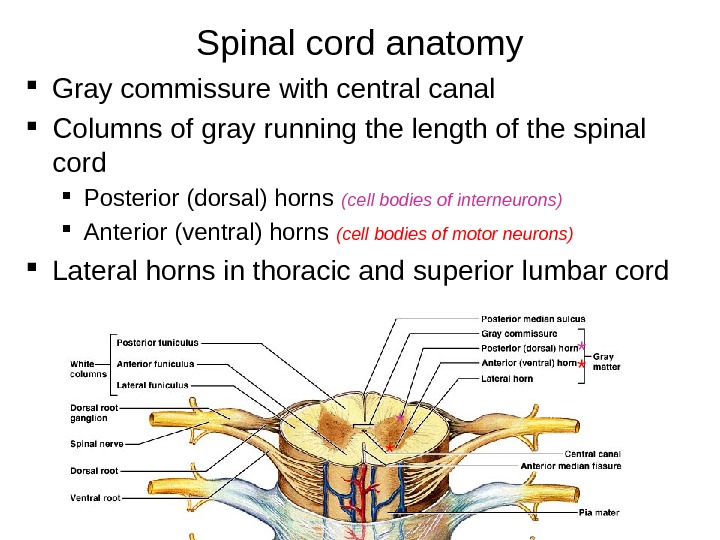
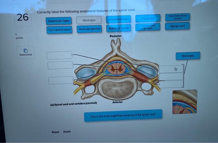
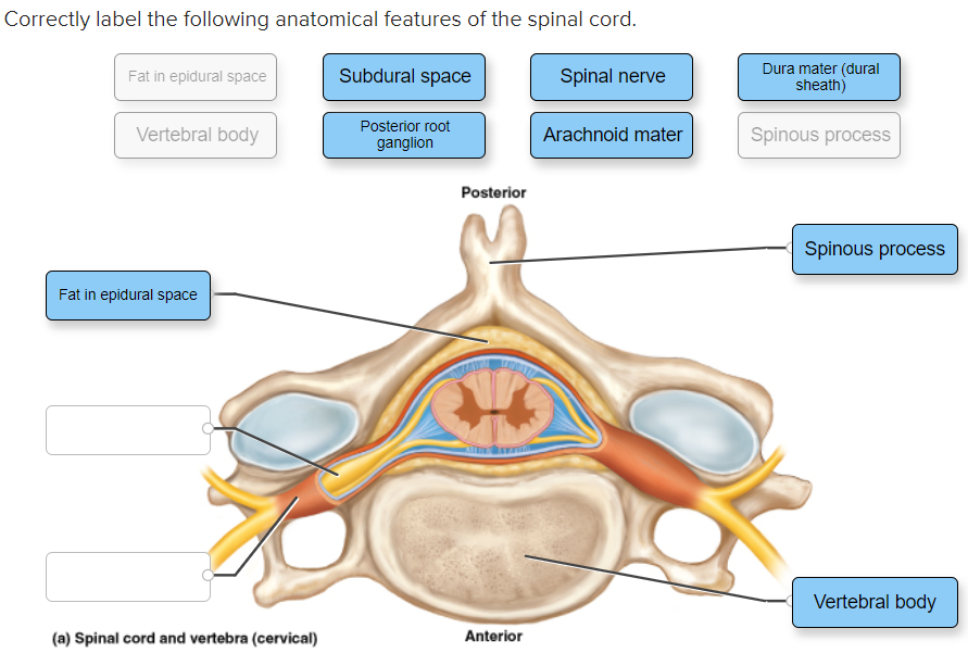
Body Cavities and Membranes: Labeled Diagram, Definitions The cross-section illustrates as if we are looking down at the spinal cord, and it shows the layers of the spinal cavity discussed above. The spinal cord shown in red is in the center of the spinal cavity. The spinal cavity is enclosed by the vertebral column shown in green. The 3 meningeal layers line the spinal cavity and are labeled by the ... (PDF) Stroke: Classification and diagnosis - ResearchGate 10/01/2018 · Studies have shown the role of imatinib in modulating the pathophysiological state of a number of disorders affecting brain and spinal cord such as Alzheimer's disease, Parkinson's disease, stroke ... Correctly label the following anatomical features of the spinal cord ... Correctly label the following anatomical features of the spinal cord. Fat in epidural space Subdural space Spinal nerve Dura mater (dural sheath) Vertebral body Posterior root ganglion Arachnoid mater Spinous process Posterior Spinous process Fat in epidural space Vertebral body (a) Spinal cord and vertebra (cervical) Anterior Apr 11 2022 05:44 AM Solved Correctly label the following anatomical features of - Chegg question: correctly label the following anatomical features of the spinal cord. 26 pimate dura materidura shout arachnoid mater meninges spinal cord farinebidural space derttelaments subdural cu ganglion 1 points posterior references meninges anterior (*) spinal cord and wertebra (cervical this is the most superficial covering of the spinal cord …
General Aspects of Traumatic Neural Diseases and Requirements ...
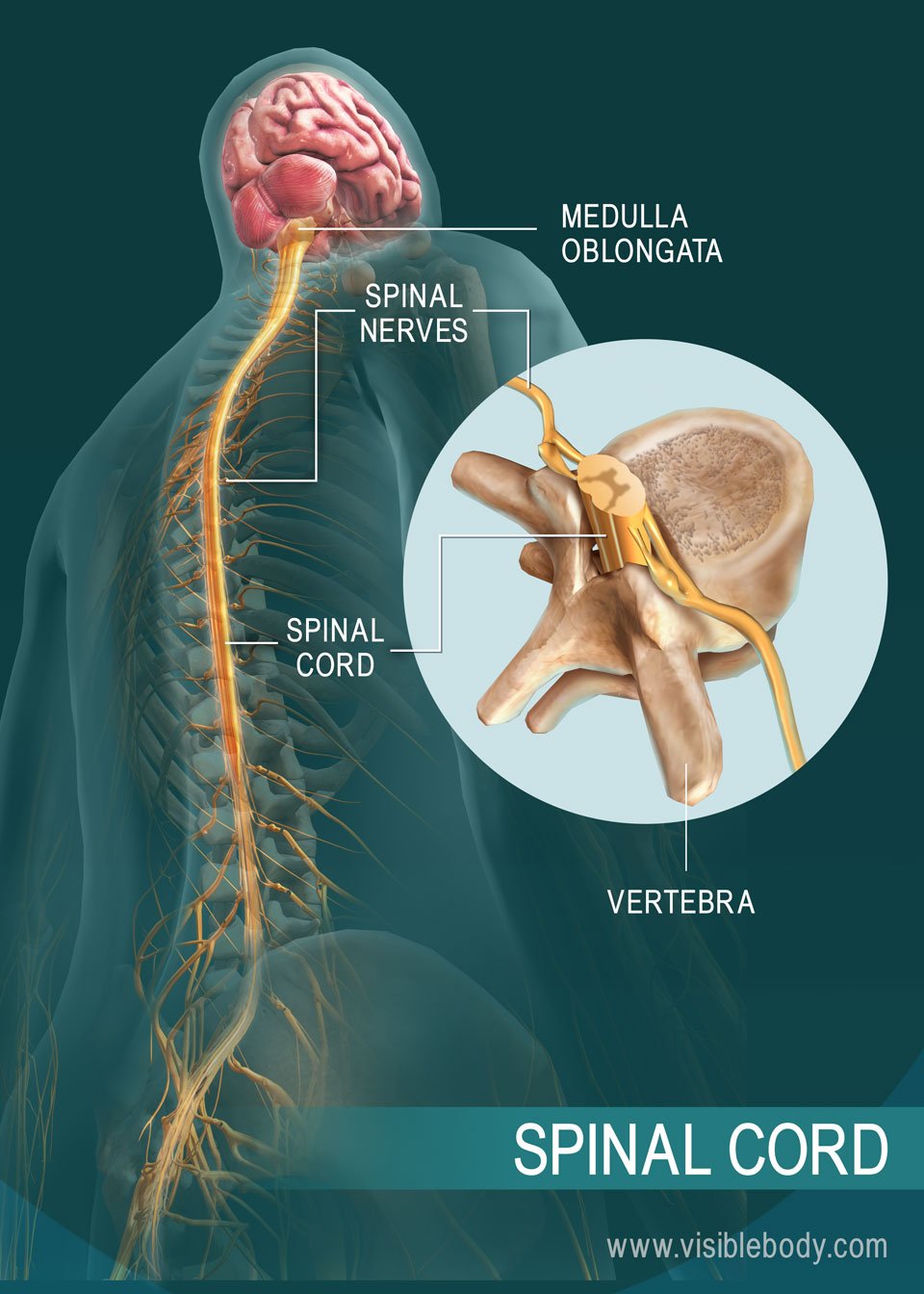
Spine Structure & Function: Parts & Segments, Spine ... - Cleveland Clinic Spinal cord and nerves: The spinal cord is a column of nerves that travels through the spinal canal. The cord extends from the skull to the lower back. Thirty-one pairs of nerves branch out through vertebral openings (the neural foramen). These nerves carry messages between the brain and muscles.

Anatomy Exam 3 study guide Flashcards | Quizlet
Success Essays - Assisting students with assignments online Get 24⁄7 customer support help when you place a homework help service order with us. We will guide you on how to place your essay help, proofreading and editing your draft – fixing the grammar, spelling, or formatting of your paper easily and cheaply.
Central Nervous System: “CNS” Prepared b y Alexey
Solved Sectional Anatomy of the Spinal Cord Correctly label - Chegg Answer... This is cross section of the vertebrae and the spinal cord Spinal cord The spinal cord is the long cord that is made up of the neural tissues and it extends from the lower part of the brain stem to the lumbar vertebrae of the human and spin … View the full answer
human skeleton - Hands and feet | Britannica
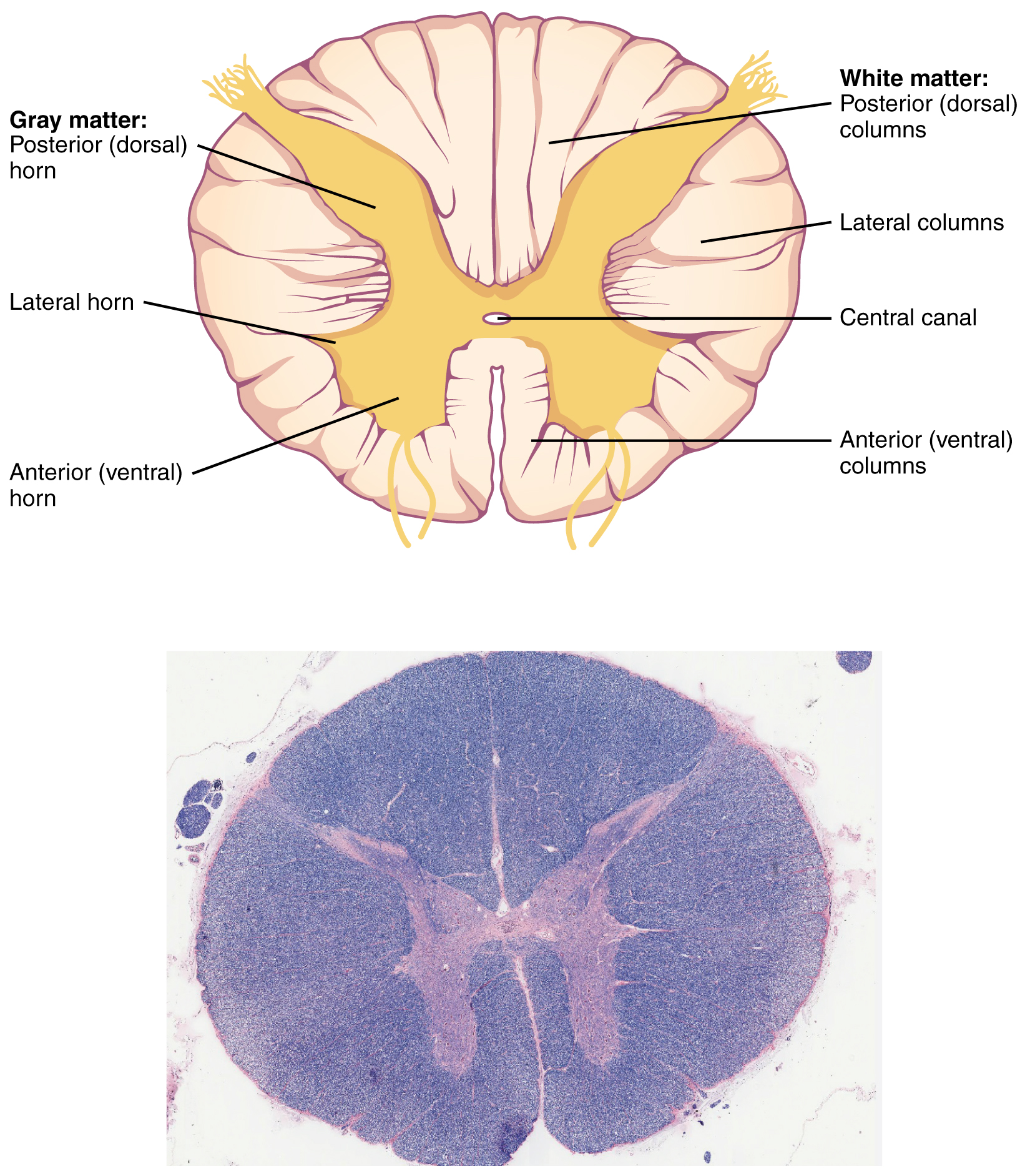
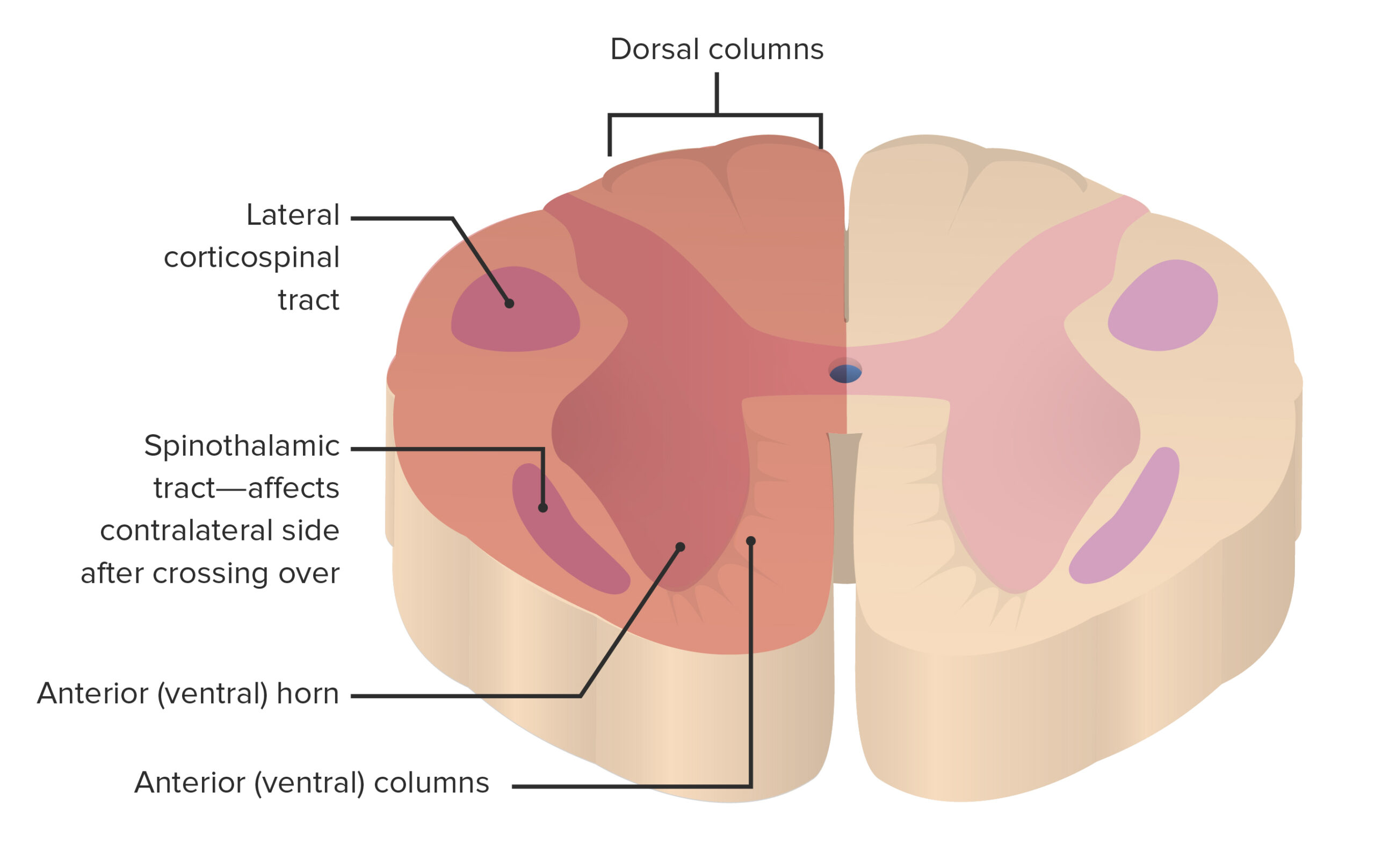
Spinal cord: Anatomy, structure, tracts and function | Kenhub The spinal cord is made of gray and white matter just like other parts of the CNS. It shows four surfaces: anterior, posterior, and two lateral. They feature fissures (anterior) and sulci (anterolateral, posterolateral, and posterior). The gray matter is the butterfly-shaped central part of the spinal cord and is comprised of neuronal cell bodies.
BrainMind.com
Spinal cord: Anatomy, functions, and injuries - Medical News Today A guide to the spinal cord: Anatomy and injuries. The spinal cord is a long bundle of nerves and cells that extends from the lower portion of the brain to the lower back. It carries signals ...
Understanding Lower Back Anatomy
Understanding Spinal Anatomy: Regions of the Spine - Cervical, Thoracic ... Cervical Spine The neck region of the spine is known as the Cervical Spine. This region consists of seven vertebrae, which are abbreviated C1 through C7 (top to bottom). These vertebrae protect the brain stem and the spinal cord, support the skull, and allow for a wide range of head movement. The first cervical vertebra (C1) is called the Atlas.
Duke Neurosciences - Lab 2: Spinal Cord & Brainstem: Surface ...
Chapter 24 Digestive System Flashcards - Quizlet 5-Distension of the rectum by feces stimulates parasympathetic reflexes. Action potentials are propagated to the defecation reflex center located in the spinal cord. 6-Action potentials stimulate contraction of the colon and rectum and relaxation of the internal anal sphincter. 7-Action potentials from the brain control the external anal sphincter.
Journal of Stem Cell Research Glial Cells Form an Integral ...
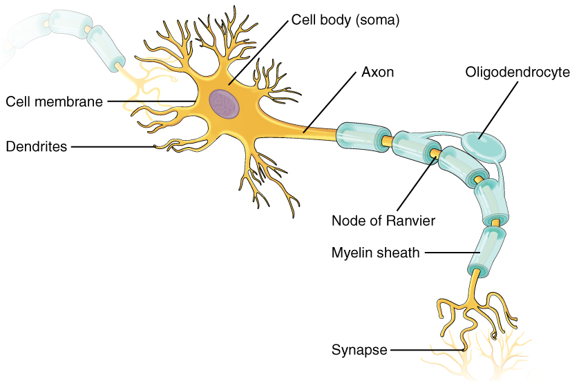
PDF Biology 201: Organization of the Nervous System 1) Use the following ... Spinal Cord Brain A) Brain B) Spinal Cord ... Source Lesson: The Nervous System: Anatomical Features & Functions 2) Label the structures of the neuron using the following terms: Myelin sheath Nucleus
How Much Computational Power Does It Take to Match the Human ...
Chapter 14 Question Set Flashcards - Quizlet Correctly identify the function of each structure that comprises a tendon reflex by dragging the appropriate label into place. Label the structures of the spinal cord. Label the spinal cord meninges and spaces. Label the white and gray matter components in the figure. Label the primary nerves of the lumbar plexus.

Polysialic-Acid-Based Micelles Promote Neural Regeneration in ...
Unit 6 Flashcards | Quizlet Label the coronal view of the head based on the hints provided. ... The micturition reflex involves impulses from the bladder traveling to which region of the spinal cord? ... Place the following anatomical structures in order to identify the correct sequence that food would pass through the body starting with ingestion. 1. Oral fissure

F21F0CF9-8C36-45E3-A106-ED46F08D7C9F.jpeg - correctly label ...
Free Science Flashcards about ANP1040 Exam 3 - StudyStack ANP1040 Exam 3. Question. Answer. Correctly label the following anatomical features of a vertebra. Vertebral arch, Spinous Process, Nucleus Pulposus, Transvere Process, Body, Vertebral Foramen, Anulous Fibrous. Correctly identify the bones and anatomical features of the bones of the skull. Frontal Bone, Maxilla, Mandible, Zygomatic Bone ...
1.3 Anatomy of the Nervous System – neurosciencecdn2
AHCDW9Notes33.pdf - 33. Award: 10.00 points Problems ... - Course Hero Correctly label the following anatomical features of the spinal cord. Explanation: The spinal cord is wrapped in a threelater protective covering called the meninges. In a crosssectional view, one can also contrast the white matter to the gray matter of the spinal cord.

Chapter 13 Worksheet Flashcards | Quizlet
chegg anatomy and physiology Solved: Cross-sectional Anatomy → Head & Neck-) Level 4 He... | Chegg.com . anatomy cross sectional neck level head. Solved: Correctly Label The Following Anatomical Features | Chegg.com . label correctly anatomical following features spinal cord anterior hom nerve mater arachnoid posterior horn transcribed text prev
JCM | Free Full-Text | Novel Therapies for the Treatment of ...
Chapter 13 Worksheet Flashcards - Quizlet Correctly label the following anatomical features of a nerve. Correctly identify and label the structures associated with the rami of the spinal nerves. Correctly identify and label the spinal nerves and their plexuses. Correctly match the nerve plexus with the spinal nerves that comprise it.
Chapter 12-Neural Tissue Flashcards - Easy Notecards
What Are The 5 Sections Of The Spine? Spinal Column Anatomy The length of the spinal cord is approximately 45 cm in men and 43 cm in women. The diameter ranges from 13 mm in the cervical and lumbar regions to 6.4 mm in the thoracic area. The cord is protected within the spinal canal and runs from the brainstem to the lumbar area where the cord fibres separate.

Biochemical and structural basis of the passive mechanical ...
Fountain Essays - Your grades could look better! We offer assignment help in more than 80 courses. We are also able to handle any complex paper in any course as we have employed professional writers who are specialized in different fields of study. From their experience, they are able to work on the most difficult assignments. The following are some of the course we offer assignment help in ...
Solved Correctly label the following anatomical features of ...
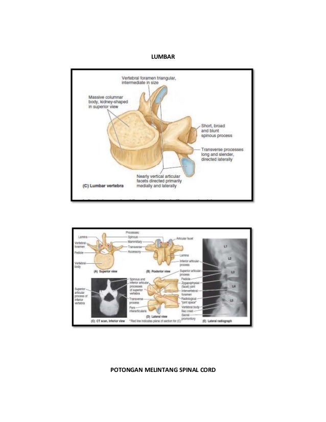
PDF We're Dream-Developers | Linn-Benton Community College cases.) Also use the key letters to correctly identify the vertebral areas in the diagram. a. b. c. body intervertebral foramina lamina d. pedicle e. spinous process g. h. transverse process vertebral arch vertebral foramen and cavity enclosing the spinal cord 2. weight-bearing portion of the vertebra 3. provide levers against which muscles ...
Unit 11 Cranial Nerves, Spinal Cord, and Reflexes
AHCDW9Notes34.pdf - 34. Award: 10.00 points Problems?... Correctly label the following anatomical features of the spinal cord. Explanation: The spinal cord is wrapped in a threelater protective covering called the meninges. In a crosssectional view, one can also contrast the white matter to the gray matter of the spinal cord.

PPT - BIOMEDICAL INSTRUMENTATION PowerPoint Presentation ...
Spinal Anatomy | Vertebral Column - SpineUniverse The Atlas is the first cervical vertebra and therefore abbreviated C1. This vertebra supports the skull. Its appearance is different from the other spinal vertebrae. The atlas is a ring of bone made up of two lateral masses joined at the front and back by the anterior arch and the posterior arch. Photo Courtesy of: SpineUniverse.com.

Descending pathways. - ppt video online download
Correctly Label the Anatomical Features of a Neuron Anatomy of the brain and spinal cord are the most obvious parts of the brain. The nucleus of a neuron is a large, round organ that contains masses of chromatin. The nucleus is also the cell's main organ, and contains the axon and deoxyribonucleic acid (DNA). The axon is the source of the signal in the neuron.

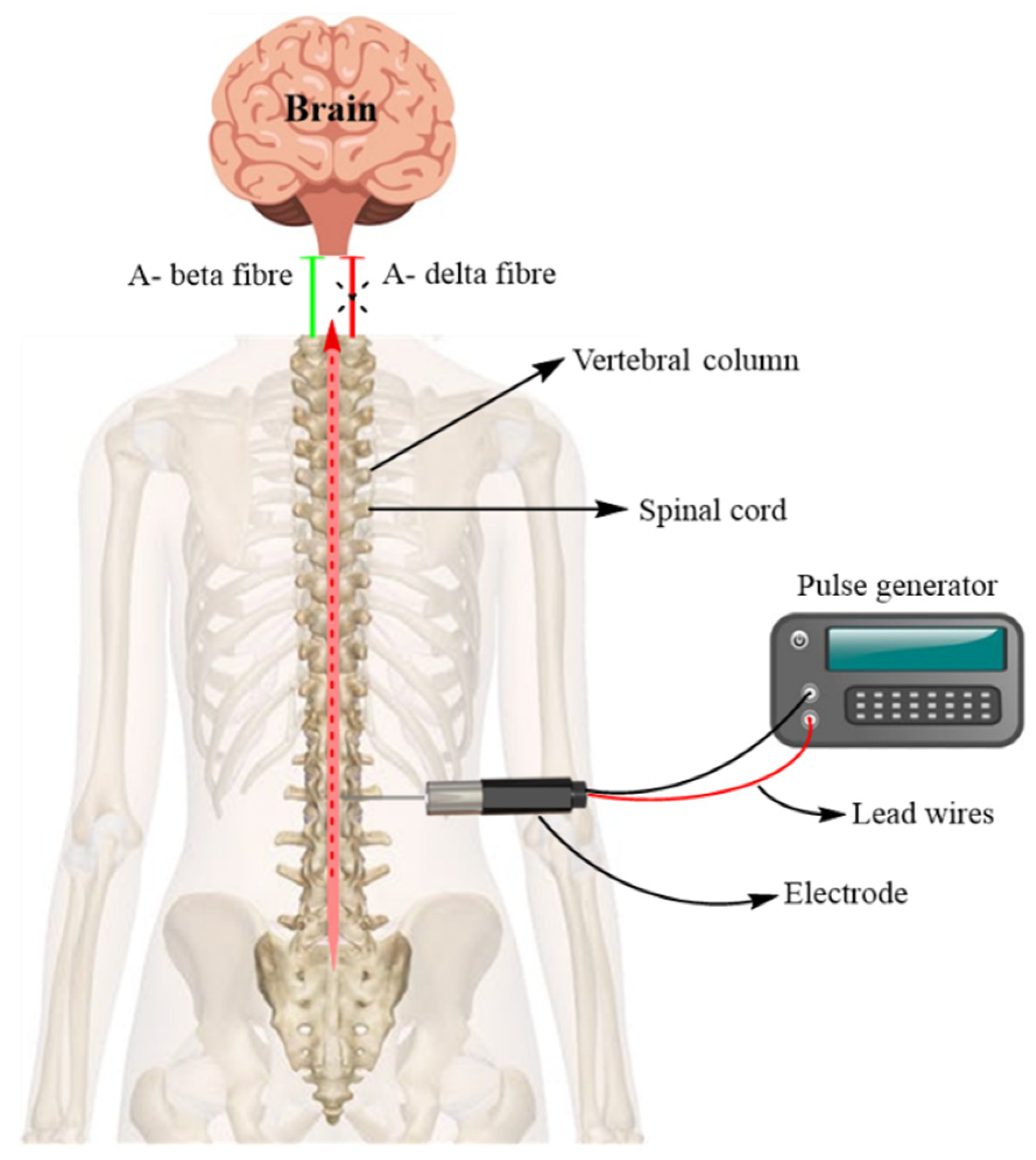
Epidural spinal cord stimulation as an intervention for motor ...
The deep crease on the anterior surface of the spinal cord is the (a ... Correctly Label The Following Anatomical Features Of The Spinal Cord. Lateral Funiculus Posterior Root Of Spinal Nerve Posterior Funiculus Posterior Horn Anterior Median Fissure Spinal Nerve Gray Commissure Spinal Nerve (B) Spinal Cord And...

What Is a Neuron? Diagrams, Types, Function, and More
Spondilitis tb

Chapter 13 Worksheet Flashcards | Quizlet

Spinal Anatomy Center | Cervical, Thoracic, and Lumbar Spine Info

25B0CC8B-E772-4BFA-B379-ABCDCE963642.jpeg - Correctly label ...

Facet Joints of the Spine's Anatomy
Spinal Cord Quiz: Cross-Sectional Anatomy | GetBodySmart
Frontiers | How Does the Central Nervous System for Posture ...

Anatomical plane - Wikipedia

A&P 1 Lab 9 Flashcards | Quizlet

230 Anatomy ideas | anatomy, anatomy and physiology, human ...
Nervous System Overview

Epidural Anesthesia and Analgesia - NYSORA | NYSORA
Solved Correctly label the following anatomical features of ...
Nervous system: Structure, function and diagram | Kenhub

AHCDW9Notes33.pdf - 33. Award: 10.00 points Problems? Adjust ...

Vertebrae - an overview | ScienceDirect Topics

Chapter 13 Worksheet Flashcards | Quizlet
Anatomy – Online USMLE Prep Course | Start now with Lecturio!
Solved Correctly label the following anatomical features of ...

Anatomical study of the medial branches of the lumbar dorsal ...
Post a Comment for "42 correctly label the following anatomical features of the spinal cord."