43 labeled microscope drawing
Light Microscope Labeled - how scanning electron microscopes work ... Light Microscope Labeled - 16 images - senior biology cell theory microscopy, what is a light microscope with pictures, 29 you will love labeling a compound microscope db, microscope imaging station gallery, Parts of the Microscope with Labeling (also Free Printouts) Parts of the Microscope with Labeling (also Free Printouts) A microscope is one of the invaluable tools in the laboratory setting. It is used to observe things that cannot be seen by the naked eye. Table of Contents 1. Eyepiece 2. Body tube/Head 3. Turret/Nose piece 4. Objective lenses 5. Knobs (fine and coarse) 6. Stage and stage clips 7. Aperture
Pseudostratified Columnar Epithelium under a Microscope with a Labeled ... Pseudostratified columnar cells labeled diagram and drawing. You already got different labeled diagrams on the pseudostratified cells. Again, I will show you a hand drawing pseudostratified columnar cell. Here, in the pseudostratified columnar hand drawing image, all structures like cells, nucleus, goblet cells, basal cells, and cilia are ...

Labeled microscope drawing
Scanning Electron Microscope (SEM) - Diagram, Working Principle ... Definition Scanning electron microscope is a classification of electron microscope that uses raster scanning to produce the images of a specimen by scanning using a focused electron beam on the surface of the specimen. An SEM creates magnified images of the specimen by probing along a rectangular area of the specimen with a focused electron beam. Microscopy: History, Types of Microscope, Diagram - Embibe A microscope known as a microscopy instrument is a device that magnifies pictures of tiny objects. Learn Microscopy history, diagrams, types, and parts. ... Diagram of Compound Microscope. Practice 11th CBSE Exam Questions. ... The cells or parts of cells are labelled with fluorescent dyes. 3. This method is used in modern life science studies ... Microscope: Types of Microscope, Parts, Uses, Diagram - Embibe There microscope anatomy includes three structural parts, i.e. head, base, and arm. Head - This is also known as the body; it carries the optical parts in the upper part of the microscope.. Base - It acts as microscopes support.It also carries microscopic illuminators. Arms - The microscope arm connects the base and the head and the eyepiece tube to the microscope base.
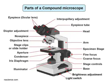
Labeled microscope drawing. Testes: Anatomy, definition and diagram | Kenhub Testis. 1/5. The testes (testicles) are male reproductive glands found in a saccular extension of the anterior abdominal wall called the scrotum. They are in ovoid shape, sized four to six centimeters in length. Testes develop retroperitoneally on the posterior abdominal wall and descend to scrotum before birth. Light Microscope- Definition, Principle, Types, Parts, Labeled Diagram ... Parts of a microscope with functions and labeled diagram 22 Types of Spectroscopy with Definition, Principle, Steps, Uses History of Microbiology and Contributors in Microbiology Microbiology of extreme environments (Types and Examples) Dark-Field Light Microscope Simple Squamous Epithelium under a Microscope with a Labeled Diagram ... Simple squamous epithelium under microscope labeled in renal corpuscle The cortex of a kidney consists of renal corpuscles and the convoluted tubule, straight tubules, nephrons, connecting tubules, and collecting ducts. You will find the medullary ray in the medulla of the kidney that comprises straight tubules and collecting ducts. Binocular Microscope Anatomy - Parts and Functions with a Labeled Diagram Now, I will discuss the details anatomy of the light compound microscope with the labeled diagram. Why it is called binocular: because it has two ocular lenses or an eyepiece on the head that attaches to the objective lens, this ocular lens magnifies the image produced by the objective lens. Binocular microscope parts and functions
Blood Histology Slides with Description and Labeled Diagram The blood is a specialized connective tissue that is fluid and circulates through the vascular channel. In the blood histology slide, you will find different types of cells with their specific features. This might be a short article where I will show you all the cells from the blood microscope slide with a labeled diagram and actual pictures. Compound Microscope- Definition, Labeled Diagram, Principle, Parts, Uses The naked eye can now view the specimen at magnification 400 times greater and so microscopic details are revealed. Alternatively, the magnification of the compound microscope is given by: m = D/ fo * L/fe where, D = Least distance of distinct vision (25 cm) L = Length of the microscope tube fo = Focal length of the objective lens Euglena Under Microscope Labeled - microscope euglena diagram ... Here are a number of highest rated Euglena Under Microscope Labeled pictures upon internet. We identified it from honorable source. Its submitted by paperwork in the best field. We acknowledge this kind of Euglena Under Microscope Labeled graphic could possibly be the most trending topic later we allowance it in google pro or facebook. Neuron under Microscope with Labeled Diagram - AnatomyLearner But, first, let's try to identify the following features from a neuron with the help of a labelled diagram. Cell body or perikaryon of a neuron Nucleus, cytoplasm, the plasma membrane of a neuron Nissl bodies in the cell body of a neuron An initial segment of axon and axon hillock Dendrites and axons of a neuron Axolemma and myelin sheath
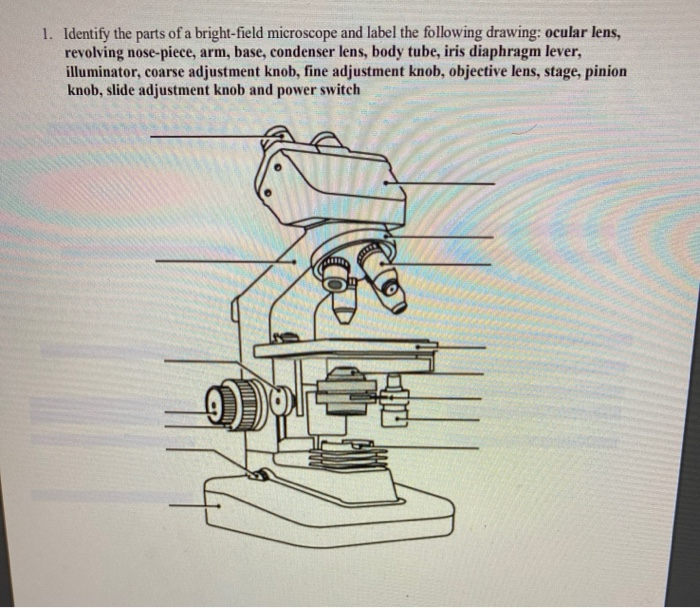
Microscope Types (with labeled diagrams) and Functions Simple microscope labeled diagram Simple microscope functions It is used in industrial applications like: Watchmakers to assemble watches Cloth industry to count the number of threads or fibers in a cloth Jewelers to examine the finer parts of jewelry Miniature artists to examine and build their work Also used to inspect finer details on products Microscope Diagram Worksheet - The Microscope Create A Labelled Diagram ... Write the letter on the line that represents each part of the microscope. Using the terms listed below, label the microscope diagram. When you can identify a part of the microscope place the . There is a printable worksheet available for download here so you can take the . Use the words from this word list to identify the parts of the microscope. Bright-field microscope (Compound light microscope) - Diagram (Parts ... Bright-field microscope parts (Labeled Diagram) Ocular Lens This microscope has two eye lenses or ocular lens on the top of the microscope that are used to focus the image from the objective lens. It is from these lenses that we see the magnified image of the specimen. Objective Lens Compound Light Microscope Diagram Worksheet - Google Groups Study manual following chapter which describes features of the initial light microscope and the function of each carbon the diagram of the microscope below. You will label sketches to compound light microscope worksheet may want to your students to use worksheets to. On a typical student compound light microscope there are 3-4 of objective lenses.
microscopy how a microscope works magnification calculations ...
Electron Microscope- Definition, Principle, Types, Uses, Labeled Diagram Electron Microscope is in the form of a tall vacuum column that is vertically mounted. It has the following components: 1. Electron gun The electron gun is a heated tungsten filament, which generates electrons. 2. Electromagnetic lenses The condenser lens focuses the electron beam on the specimen.

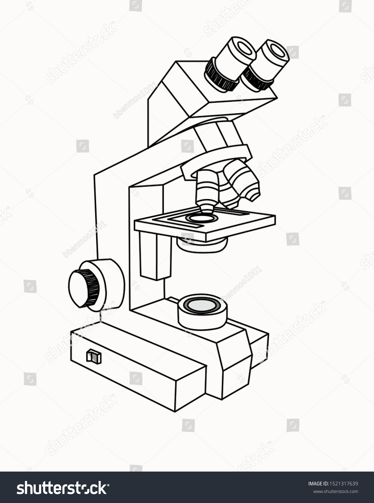
Old microscope color sketch engraving vector illustration ...
Simple Microscope - Parts, Functions, Diagram and Labelling Simple microscope - It was the first microscope ever created. It was created by Antony van Leeuwenhoek in the 17th century. He combined a convex lens and a holder for specimens. It looks like a magnifying glass because it has the ability to magnify between 200 and 300 times.

Unlabeled Microscope Diagram posted by Christopher Thompson
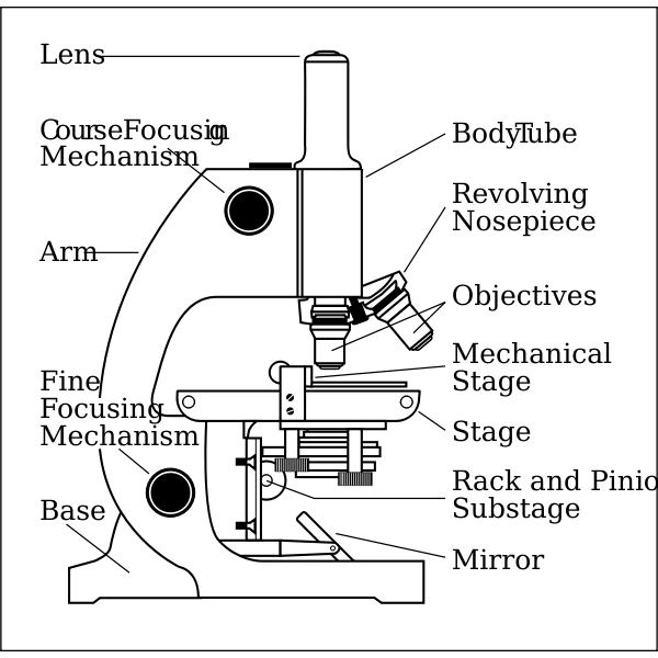
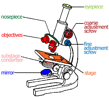
Parts of a microscope with functions and labeled diagram - Microbe Notes Figure: Diagram of parts of a microscope There are three structural parts of the microscope i.e. head, base, and arm. Head - This is also known as the body. It carries the optical parts in the upper part of the microscope. Base - It acts as microscopes support. It also carries microscopic illuminators.

File:Labelledmicroscope.gif - Wikimedia Commons
Compound Microscope - Diagram (Parts labelled), Principle and Uses See: Labeled Diagram showing differences between compound and simple microscope parts Structural Components The three structural components include 1. Head This is the upper part of the microscope that houses the optical parts 2. Arm This part connects the head with the base and provides stability to the microscope.
Microscope side vector drawing with parts labelled | Free SVG
Microscope Parts, Function, & Labeled Diagram - slidingmotion Microscope parts labeled diagram gives us all the information about its parts and their position in the microscope. Microscope Parts Labeled Diagram The principle of the Microscope gives you an exact reason to use it. It works on the 3 principles. Magnification Resolving Power Numerical Aperture. Parts of Microscope Head Base Arm Eyepiece Lens
Color the Microscope Parts
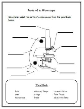
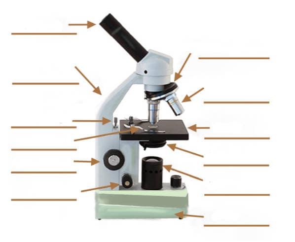
Label The Parts Of A Microscope Worksheet Answers You can use the word bank below to fill in the blanks or cut. Label the parts of a microscope worksheet answers. Students label the parts of the microscope in this photo of a basic laboratory light microscope. Files include a link to editable doc so you can rewrite a. Power 10 x 4 40 Power 10 x 10 100 Power 10 x 40 400 What happens as the power ...

Mirror microscope Vector Art Stock Images | Depositphotos
Inverted Microscope- Definition, Principle, Parts, Labeled Diagram ... invented in 1850 by a faculty member of medical college of louisiana, named j. lawrence smith, this microscope just like it sounds is a light microscope that has its components placed in an inverted order, this means, light source and condenser lens are placed above the specimen stage, pointing down, while the objectives and the turret are found …
Microscope With Labels clip art Vectors graphic art designs ...
Microscope- Definition, Parts, Functions, Types, Diagram, Uses It is a type of fluorescence microscope that is used to produce 2-D or 3-D images of relatively thick specimens. In this type, the excitation light is focused on a specific spot of sample lying on the focal plane. The focus spot is optically manipulated to scan the entire sample and generate a 3-D image.
Microscope | Other Quiz - Quizizz
Microscope, Microscope Parts, Labeled Diagram, and Functions Multiply the magnification of the eyepiece (ocular lens) by the magnification of the objective lens in use to calculate the total magnification of any object viewed under the microscope. This can be demonstrated using the formula. Total magnification = ocular lens x objective lens
Glossary of terms used in microscopy – Quekett Microscopical Club
Simple Microscope - Diagram (Parts labelled), Principle, Formula and Uses The working principle of a simple microscope is that when a lens is held close to the eye, a virtual, magnified and erect image of a specimen is formed at the least possible distance from which a human eye can discern objects clearly. Magnification formula The magnification power of a simple microscope is expressed as: M = 1 + D/F Where
Compound Microscope Drawing - ClipArt Best - ClipArt Best ...
Microscope: Types of Microscope, Parts, Uses, Diagram - Embibe There microscope anatomy includes three structural parts, i.e. head, base, and arm. Head - This is also known as the body; it carries the optical parts in the upper part of the microscope.. Base - It acts as microscopes support.It also carries microscopic illuminators. Arms - The microscope arm connects the base and the head and the eyepiece tube to the microscope base.
Label the Microscope Parts for Elementary School Students by ...
Microscopy: History, Types of Microscope, Diagram - Embibe A microscope known as a microscopy instrument is a device that magnifies pictures of tiny objects. Learn Microscopy history, diagrams, types, and parts. ... Diagram of Compound Microscope. Practice 11th CBSE Exam Questions. ... The cells or parts of cells are labelled with fluorescent dyes. 3. This method is used in modern life science studies ...

Label the microscope — Science Learning Hub
Scanning Electron Microscope (SEM) - Diagram, Working Principle ... Definition Scanning electron microscope is a classification of electron microscope that uses raster scanning to produce the images of a specimen by scanning using a focused electron beam on the surface of the specimen. An SEM creates magnified images of the specimen by probing along a rectangular area of the specimen with a focused electron beam.

Compound Microscope Parts – Labeled Diagram and their ...
Laboratory microscope sketch Stock Vector Image & Art - Alamy

Compound Microscope Parts, Functions, and Labeled Diagram ...

Free Microscope Drawing, Download Free Microscope Drawing png ...

Label Microscope Diagram - EnchantedLearning.com

Draw a well labelled diagram of a microscope. - Brainly.in
Compound Microscope Parts – Labeled Diagram and their ...

Types of Microscopes

Parts of a Microscope - SmartSchool Systems

Microscope Labeling Diagram | Quizlet

Biology Notes for A level: #75 Drawings
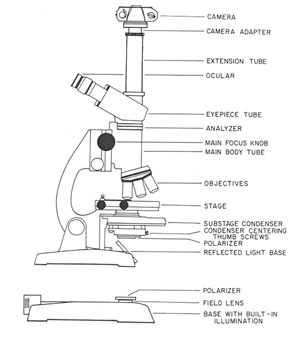
The Microscope

Free Microscope Drawing, Download Free Microscope Drawing png ...
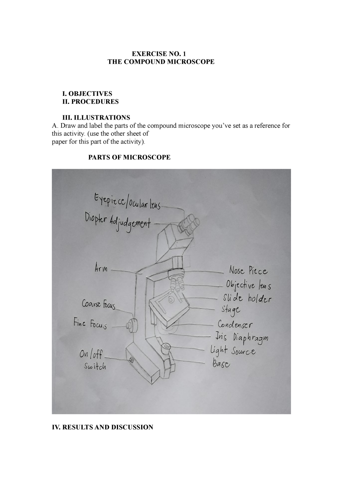
Compound microscopic microbiology - EXERCISE NO. 1 THE ...

How To Draw A Microscope, Step by Step, Drawing Guide, by ...
Parts of a microscope with functions and labeled diagram

Cartoon Sticker Stick Icon Decal Label Microscope Science ...

Label microscope pt.1 Diagram | Quizlet

microscope drawing | Clipart Panda - Free Clipart Images
Parts of the Microscope with Labeling (also Free Printouts ...

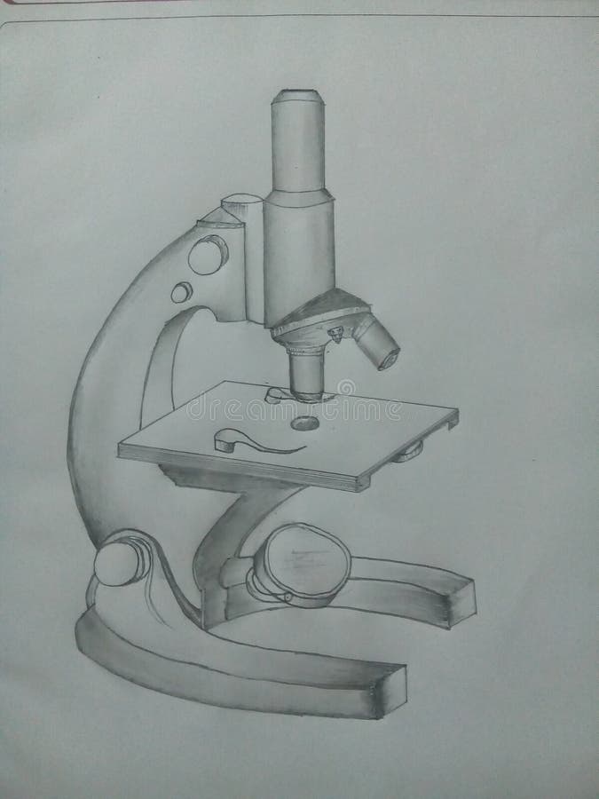
How to Draw a Microscope - Really Easy Drawing Tutorial
Solved 7. The Microscope 1. In a compound microscope: a. The ...
Vektor Stok Vector Microscope (Tanpa Royalti) 1209424708 ...
Compound Microscope Stock Illustrations – 727 Compound ...

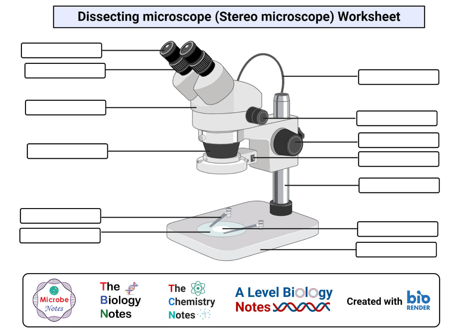
Parts of Stereo Microscope (Dissecting microscope) – labeled ...
Draw a diagram of compound microscope and electrone ...

Biology 4 U on Twitter: "Try this labelled diagram Quiz on ...
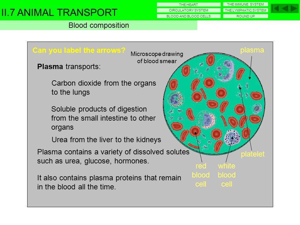
Microscope drawing of blood smear - ppt video online download

22 Parts Of a Microscope With Their Function And Labeled ...

How to Draw a Cartoon Microscope – Draw Swan
Post a Comment for "43 labeled microscope drawing"