43 labeled gel electrophoresis photo
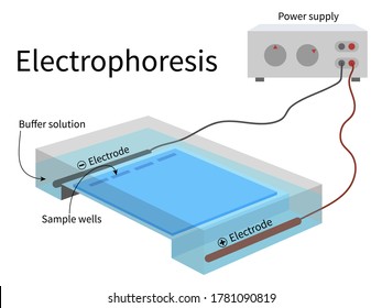
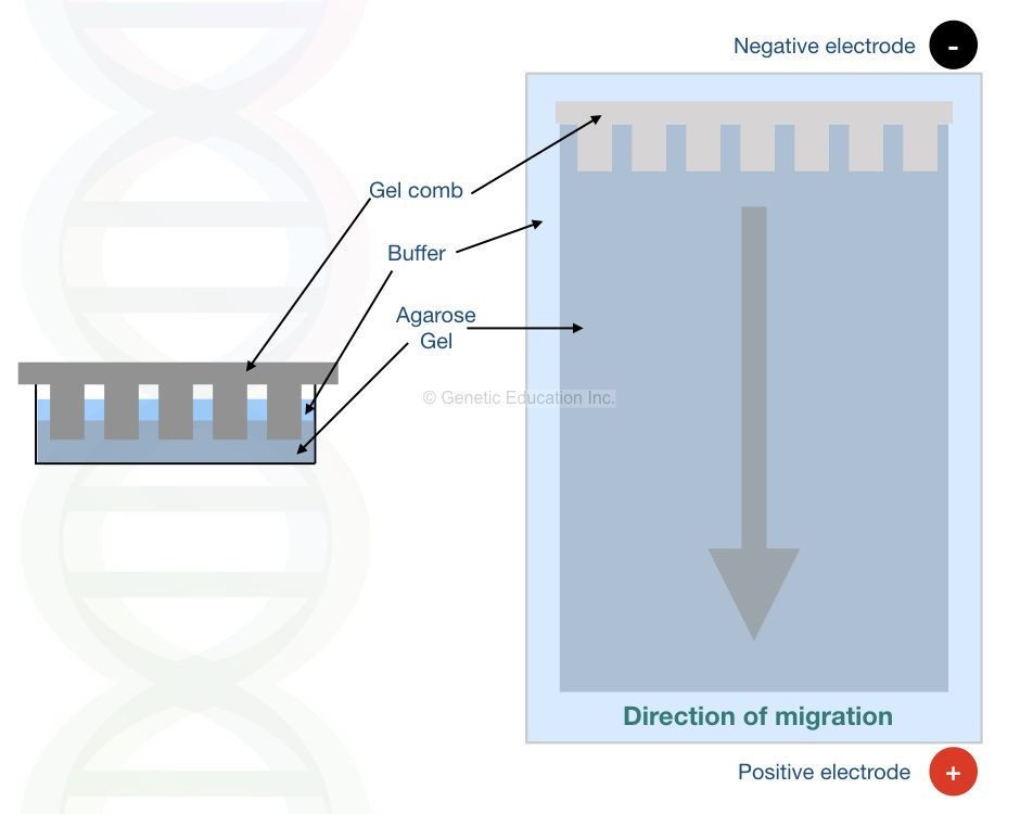
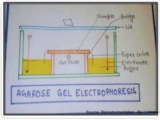
Part 2: Analysing and Interpreting (Agarose) Gel Electrophoresis Results The agarose gel electrophoresis is a molecular genetic technique used to separate DNA on the basis of size and charge of it. The negatively charged DNA migrates towards the positive node under the influence of the current. The results of agarose electrophoresis are affected by some of the factors enlisted below, The concentration of gel How to Interpret DNA Gel Electrophoresis Results | GoldBio Agarose gel electrophoresis is a molecular biology method to analyze and separate DNA fragments based on their size. When you use gel electrophoresis to help you with molecular cloning, you may run into a common problem. For an example, you are ready to excise your digested plasmid DNA from agarose.
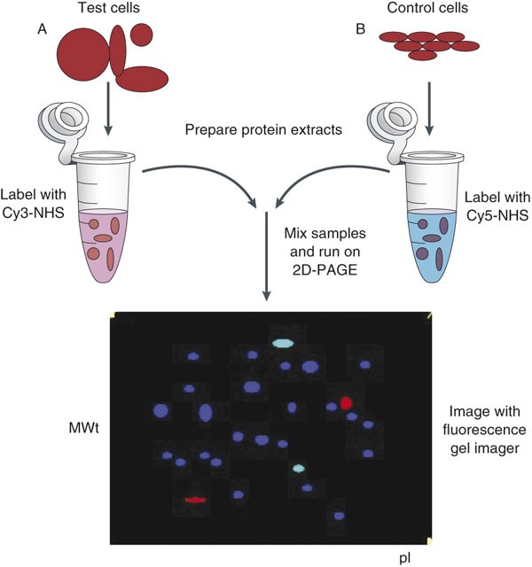
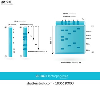
Gel electrophoresis: Types, introduction and their applications Gel electrophoresis contains supporting medium as gel and it is of various types as shown below-Paper gel electrophoresis,agarose gel electrophoresis. ... Up to 3 different protein samples can be labeled with size and charge matched fluorescent dyes (for example Cy3, Cy5, Cy2) the three samples are mixed, loaded and 2D electrophoresis is ...

Labeled gel electrophoresis photo
Gel Electrophoresis: Molecular Biology Science Activity | Exploratorium ... Prepare your gel: Make a 0.2% sodium bicarbonate buffer by dissolving 2 grams of baking soda in 1 liter of water. You will need approximately 100 milliliters per set up—half to make the gel and half to run your samples. Make a 1% gel solution by adding 0.5 g of agar-agar powder to 50 mL of sodium bicarbonate buffer. How does qPCR work (technology basics)? - BioSistemika Figure 6: Shows a picture (negative) of an agarose gel after electrophoresis, stained with ethidium bromide and visualized under UV light. Columns ‘M’ contain molecular weight markers, each band representing a DNA fragment of a known length -shortest being on the bottom of the gel and longest being on the top of the gel. Droplet-based microfluidics - Wikipedia Surfactants play an important role in droplet-based microfluidics. The main purpose of using a surfactant is to reduce the interfacial tension between the dispersed phase (droplet phase, typically aqueous) and continuous phase (carrier liquid, typically oil) by adsorbing at interfaces and preventing droplets from coalescing with each other, therefore stabilizing the droplets in a stable ...
Labeled gel electrophoresis photo. What is gel electrophoresis? - YourGenome To make a gel, agarose powder is mixed with an electrophoresis buffer and heated to a high temperature until all of the agarose powder has melted. The molten gel is then poured into a gel casting tray and a "comb" is placed at one end to make wells for the sample to be pipetted into. Dissolved oxygen from microalgae-gel patch promotes chronic ... May 15, 2020 · Then, the gaseous-oxygen group was treated with medical grade 100% oxygen at 1.5 atm for 3 hours, while the alga-gel group was treated with alga-gel (1 × 10 9 cells/ml) into the upper chamber with NIR LED for 1 hour, followed by the use of an illumination incubator. After 24 hours, cells were fixed with 4% paraformaldehyde (PFA) for 15 min ... Fluorophore-Labeled Primers Improve the Sensitivity, Versatility, and ... Electrophoresis was carried out for 14 h at 60°C and 85 V. After electrophoresis, gels were either imaged directly or first stained with SYBR Green I (Molecular Probes, Eugene, OR) at a 1:10,000 dilution for 1 h and then destained for 15 min in 1× Tris-acetate-EDTA buffer. Gel electrophoresis — Science Learning Hub Isoelectric focusing (IEF) and agarose gel electrophoresis are two ways that proteins can be separated by their different electrical charges. Unlike SDS-PAGE, the proteins are usually kept in their native (folded) state. The type of gel that is used, and the solution around the gel, are also different. In agarose gel electrophoresis, proteins ...
Gel Electrophoresis - an overview | ScienceDirect Topics Gel electrophoresis is an analytical technique that allows size separation of DNA as well as other macromolecules. For gel electrophoresis, a DNA sample is loaded at one end of a gel matrix (usually agarose or acrylamide) that provides a uniform pore size through which the DNA molecules can move. 551 Gel Electrophoresis Premium High Res Photos - Getty Images 551 Gel Electrophoresis Premium High Res Photos Browse 551 gel electrophoresis stock photos and images available, or search for dna or pcr to find more great stock photos and pictures. Related searches: dna pcr biotechnology chromatography dna gel of 10 NEXT Two-dimensional gel electrophoresis (2D-GE) image analysis b ... - LWW With this, the proteins are separated as spots, which are revealed with stains such as Coomassie blue or silver stain, to then capture images of the gel. These images are then analyzed to identify the points and study the protein content, as well as continue with subsequent proteomic studies by other strategies. [1] Gel Electrophoresis - Dolan DNA Learning Center In the 1970s, the powerful tool of DNA gel electrophoresis was developed. This process uses electricity to separate DNA fragments by size as they migrate through a gel matrix. ... GMO gel. Gel photo of PCR amplification to detect GMO or transgenes in food. ID: 16134; Source: DNAi; 16529. Animation 24: The RNA message is sometimes edited.
Understanding and Interpreting Serum Protein Electrophoresis In zone electrophoresis, for example, different protein subtypes are placed in separate physical locations on a gel made from agar, cellulose, or other plant material. 2, 3 The proteins are ... Gel electrophoresis Images, Stock Photos & Vectors | Shutterstock Predict Help Gel electrophoresis royalty-free images 769 gel electrophoresis stock photos, vectors, and illustrations are available royalty-free. See gel electrophoresis stock video clips Image type Orientation Color People Artists More Sort by Science Biology College and University dna agarose gel electrophoresis gel electrophoresis Agarose Gel Electrophoresis - an overview | ScienceDirect Topics Agarose gel electrophoresis is widely used for separation of DNA and RNA samples in events like restriction fragment analysis, polymerase chain reaction product analysis, checking the integrity of genomic DNA, and purification of nucleic acids. DNA and RNA are negatively charged and during electrophoresis, the side of the gel having wells is ... Activity 2 - Gel Electrophoresis of Dyes - APS Home Place gel into electrophoresis unit. Add 150 ml 1X TBE buffer to completely fill the box and to cover the top gel surface with about 2 mm of buffer. Note: At this point the gel box can be covered and left until the next day if necessary. On the gel load 5-10 µl of each dye into a well.
Study the diagram given and answer the question.Identify the ...
Gel electrophoresis (article) | Khan Academy Gel electrophoresis. Gel electrophoresis. This is the currently selected item. DNA sequencing. DNA sequencing. Applications of DNA technologies. Practice: Biotechnology. Sort by: Top Voted. Gel electrophoresis. DNA sequencing. Up Next. DNA sequencing. Biology is brought to you with support from the Amgen Foundation.

Difference Gel Electrophoresis: Methods and Protocols Edition ...
E-Editor 2.0 Software | Thermo Fisher Scientific - US Analysis of E-PAGE™ gel and E-Gel® results is fast and convenient, for both stained gels and blots, using the Windows®-based E-Editor™ 2.0 software. E-Editor™ 2.0 is user-friendly software that quickly arranges and displays your electrophoresis results. Just capture an image of the gel and use the E-Editor™ 2.02 software to align and arrange the lanes in the image, save the ...
734 Gel electrophoresis Images, Stock Photos & Vectors ...
PDF Gel Electrophoresis: How Does It Work - Purdue University a. After you find out what dyes you are using, draw a picture of the gel and the wells. Label which dyes you will put in each well. b. When you load a gel, it is very important that you do not damage the gel in any way. You must be very careful not to "jab" the gel with the end of your pipet. Ideally, you shouldn't even touch the gel with the ...
Draw a diagram of a typical agarose gel electrophoresis ...
Lab #6 Gel Electrophoresis and Nanodrop Spectrophotometry Add a Low mass standard to the labeled well. Plug in the gel electrophoresis box and run at 100v for ~20-30 minutes. ( You will know if it is working if you see bubbles forming in the buffer liquid) Carefully and with gloves, turn off the box and retrieve the gel. Transfer the gel into a small container and bring to the lab.
Investigations on therapeutic glucocerebrosidases through ...
Gel Electrophoresis - Definition, Purpose and Steps - Biology Dictionary The broad steps involved in a common DNA gel electrophoresis protocol: 1. Preparing the samples for running The DNA is isolated and preprocessed (e.g. PCR, enzymatic digestion) and made up in solution with some basic blue dye to help visualize the movement of the sample through the gel. 2. An agarose TAE gel solution is prepared

PDF) Difference gel electrophoresis | Kai Stühler - Academia.edu
3 Ways to Read Gel Electrophoresis Bands - wikiHow Gel electrophoresis is a type of biotechnology that separates molecules based on their size to interpret an organism's DNA. An enzyme is used to separate a strand of DNA from a source and the DNA is suspended in a dye. Then, the dye is applied to a negatively-charged gel on one side of a sheet.

Evaluation of PCR-labeled probes by agarose gel ...
What does this RNA gel electrophoresis image indicate ... - ResearchGate This is the image of Total RNA non- denaturing gel electrophoresis. The 2 rRNA bands (28s and 18s) are prominent with 2:1 intensity but there is no smearing in between them that indicates presence...
SOLVED: Take a screenshot of the labeled image of your gel ...
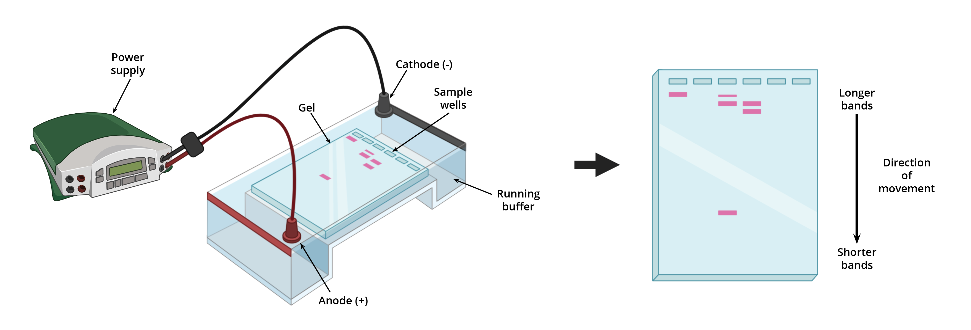
gel electrophoresis | Britannica The gel electrophoresis apparatus consists of a gel, which is often made from agar or polyacrylamide, and an electrophoretic chamber (typically a hard plastic box or tank) with a cathode (negative terminal) at one end and an anode (positive terminal) at the opposite end. The gel, which contains a series of wells at the cathode end, is placed inside the chamber and covered with a buffer solution.
Two-dimensional difference gel electrophoresis | Nature Protocols
What negative controls are typically included in ... - Qiagen 3. A no amplification control (NAC) omits the DNA polymerase from the PCR reaction. This is a control for background fluorescence that is not a function of the PCR. Such fluorescence is typically attributable to the use of a degraded, dual-labeled probe. This control is unnecessary when utilizing SYBR-Green probe chemistries.

Practical 3 part I - agarose gel electrophoresis Diagram ...
Gel Electrophoresis - University of Utah Sort and measure DNA strands by running your own gel electrophoresis experiment. See how gel electrophoresis is used in forensics. Can DNA Demand a Verdict? Try it Yourself. How to Build an Electrophoresis Chamber (PDF) Colorful Electrophoresis. Funding.
SOLVED: Take a screenshot of the labeled image of your gel ...
PDF Lab 4: Gel Electrophoresis - Vanderbilt University Gel electrophoresis is a method of separating DNA fragments by movement through a Jello-like substance called agarose. Derived from a seaweed polysaccharide, agarose gels form small pores ... terms are labeled on the gel, and the loading key is labeled according to each lane. 1000bp 500bp 2000bp 250bp 100bp Lane 4 Lane Sample 1 DNA Ladder

Figure legends Figure 1: Agarose gel electrophoresis (2 ...
How To Label Gel Electrophoresis Images / Electrophoresis - YouTube ... How to label a gel electrophoresis image. This tutorial is all about how to quickly edit & label pcr gel image using imagej software. 1.take your jpg or png file of your gel and open it with a photo editing program (gimp). How do i distinguish between dna and rna on a gel.
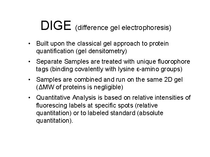
DIGE difference gel electrophoresis Built upon the classical
A complete guide for analysing and interpreting gel electrophoresis results Agarose gel electrophoresis is an important technique in molecular genetics for a long. DNA bands can only be visualized using agarose gel electrophoresis. In genomic research, analyzing and interpreting the agarose gel electrophoresis results are very crucial. A lot of expertise and experience are required for Interpreting gel electrophoresis ...
1,835 Electrophoresis Images, Stock Photos & Vectors ...
Agarose Gel Electrophoresis: Results Analysis - Study.com Gel electrophoresis is a laboratory procedure used to separate biological molecules with an electrical current. Previously, we've discussed gel electrophoresis in the context of analyzing DNA....

Given below is the diagram representing the observations made ...
Annotating A Gel | Get Your Science On Wiki | Fandom Part 1. Photo Editing: 1.Take your JPG or PNG file of your Gel and open it with a photo editing program (GIMP). 2. Under "Image" --> "Transform" rotate your picture by 90 degrees so that your wells are on top of the page. 3. Using the Crop tool Cut out the black borders leaving only the gel. 4.
Image Analysis and Pattern Extraction of Proteins Classes ...
PDF Protocol 4: Gel Electrophoresis Teacher Version the Genome PROTOCOL 4: GEL ELECTROPHORESIS TEACHER VERSION ⎕ STEP 1 Set up and turn on the LONZA system and laptop so it is ready to go once the gels are loaded. ⎕ STEP 2 Open a fresh gel cassette package from the LONZA system and insert gel cassette into gel dock by sliding into place. Then remove white seals from gel cassette.

cont.) GEL ELECTROPHORESIS (basic concept) - ppt video online ...
(MB) ASCP Practice Exam Questions Flashcards | Quizlet After an amplification procedure followed by gel electrophoresis, you took a photo of your gel. You notice consistent bands in all your gel lanes. However, you also see what appears to be a faint but noticeable band in your reagent blank lane. What is your best explanation for the reagent blank band?

Evaluation of PCR-labeled probes by agarose gel ...
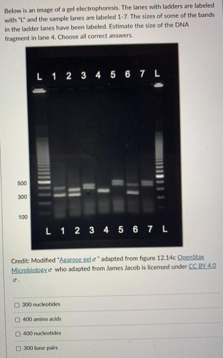
PDF Gel electrophoresis: sort and see the DNA 10. On the gel picture below, (a) circle the smallest fragment produced by a restriction enzyme and label it "smallest." (b) circle the largest fragment produced by a restriction enzyme and label it "largest." 11. In one or two sentences, summarize the technique of gel electrophoresis. Student answers DNA restriction fragment size chart

Visualizing and Characterizing DNA, RNA, and Protein ...
Droplet-based microfluidics - Wikipedia Surfactants play an important role in droplet-based microfluidics. The main purpose of using a surfactant is to reduce the interfacial tension between the dispersed phase (droplet phase, typically aqueous) and continuous phase (carrier liquid, typically oil) by adsorbing at interfaces and preventing droplets from coalescing with each other, therefore stabilizing the droplets in a stable ...
Citrate inhibition of cisplatin reaction with DNA studied ...
How does qPCR work (technology basics)? - BioSistemika Figure 6: Shows a picture (negative) of an agarose gel after electrophoresis, stained with ethidium bromide and visualized under UV light. Columns ‘M’ contain molecular weight markers, each band representing a DNA fragment of a known length -shortest being on the bottom of the gel and longest being on the top of the gel.

What is gel electrophoresis? – YourGenome
Gel Electrophoresis: Molecular Biology Science Activity | Exploratorium ... Prepare your gel: Make a 0.2% sodium bicarbonate buffer by dissolving 2 grams of baking soda in 1 liter of water. You will need approximately 100 milliliters per set up—half to make the gel and half to run your samples. Make a 1% gel solution by adding 0.5 g of agar-agar powder to 50 mL of sodium bicarbonate buffer.

Passivated gel electrophoresis of charged nanospheres by ...
Agarose Gel Electrophoresis: Definition, Principle, Process ...
Archive image from page 72 of The development of a cosmid ...
Agarose gel electrophoresis of labeled DNA in which the same ...
Gel electrophoresis

Identification of Dynamic Proteome Changes Upon Ligand ...

Two-dimensional gel electrophoresis of CyDye-labeled human ...

PPT - Gel Electrophoresis PowerPoint Presentation, free ...

A) Agarose gel electrophoresis of hybrid plasmids extracted ...
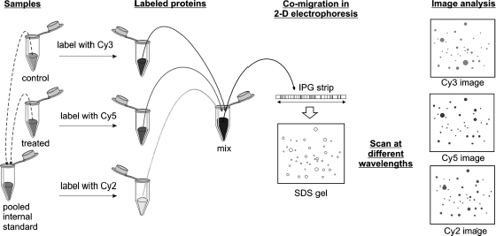
Figure 1_1_978-1-58829-679-5 | Capabilities Using 2-D DIGE in ...

Evaluation of PCR-labeled probes by agarose gel ...

InDesign Labeling / Annotating PCR Gel Pictures - Advanced Tutorial (Part 12)

How is gel electrophoresis used in agriculture? | Homework ...

File:Gelelectrophoresis.jpg - Wikimedia Commons
Archive image from page 72 of The development of a cosmid ...
PROTOCOL 4: GEL ELECTROPHORESIS

ImageJ for Editing & Labelling PCR Gel Image | Biotechnology
Agarose gel electrophoresis (AGE) image of the PCR products ...
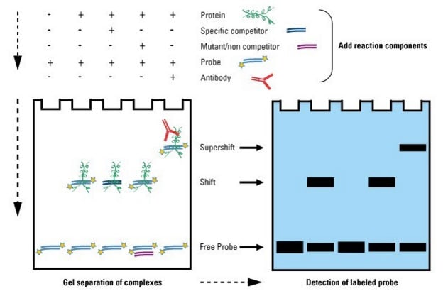
Gel Shift Assays (EMSA) | Thermo Fisher Scientific - US

View Image
Gel Electrophoresis | AAT Bioquest
Solved Below is an image of a gel electrophoresis. The lanes ...
Post a Comment for "43 labeled gel electrophoresis photo"