44 how to label gel electrophoresis images
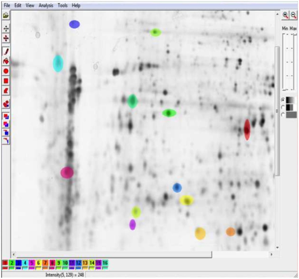
Two-Dimensional Gel Electrophoresis - an overview ... Two-Dimensional Gel Electrophoresis. Two-dimensional gel electrophoresis (2-DE) is a key tool for comparative proteomics research. In 2-DE, mixtures of proteins are separated by charge (isoelectric point, pI) in the first dimension and further separated by mass in the second dimension on 2-D gels. Goat anti-Mouse IgG (H+L) Highly Cross-Adsorbed, Alexa Fluor ... Most commonly, secondary antibodies are generated by immunizing the host animal with a pooled population of immunoglobulins from the target species and can be further purified and modified (i.e. immunoaffinity chromatography, antibody fragmentation, label conjugation, etc.) to generate highly specific reagents.
Overview of Post-Translational Modification - Thermo Fisher Scientific Samples are first reacted with MMTS to block free sulfhydryls in S-nitrosylated proteins. The S-nitrosocysteines are then selectively reduced with ascorbate for labeling with the Thermo Scientific iodoTMTzero Label Reagent. Subsequently, the supplied anti-TMT antibody is used to detect the TMT-labeled proteins in a western blot.
How to label gel electrophoresis images
ChemiDoc Imaging Systems | Bio-Rad Find the right Bio-Rad protein gel for your application. Choose SDS-PAGE and native PAGE gels, convert to TGX Precast Gels, or choose specialized gel chemistries. Image Lab Software. Image acquisition and analysis software for Bio-Rad Gel Doc, ChemiDoc, and GS-900 Systems. Capture and analyze digital image data from electrophoresis gels and blots. Activity 3: Restriction Enzyme Analysis Set up the electrophoresis apparatus as described in Gel Electrophoresis of Dyes - Activity 2. Load 20 µl of each sample into a well as shown in figure 2 above. Use the tips that were left in each tube or make sure that you use a new tip for each sample if you stored the tubes overnight. Turn on the current for about 30-45 minutes. Stain-Free Imaging Technology | Bio-Rad Images of the gel before and after transfer and of the membrane after transfer were taken using the Criterion Stain-Free Imager. Serial 1:2 dilutions of hemoglobin (starting quantity, 80 ng), with 1.8 μg of BSA/lane as a carrier (top band), were electrophoretically separated on a 4–20%, 26-well Criterion Stain-Free Gel.
How to label gel electrophoresis images. Classzone.com has been retired - Houghton Mifflin Harcourt Connected Teaching and Learning. Connected Teaching and Learning from HMH brings together on-demand professional development, students' assessment data, and relevant practice and instruction. Diagnostics | Free Full-Text | Deep Learning Assisted ... 2 days ago · Haemoglobin (Hb) electrophoresis is a method of blood testing used to detect thalassaemia. However, the interpretation of the result of the electrophoresis test itself is a complex task. Expert haematologists, specifically in developing countries, are relatively few in number and are usually overburdened. To assist them with their workload, in this paper we present a novel method for the ... Primer3 Input (version 0.4.0) Pick left primer, or use left primer below: Pick hybridization probe (internal oligo), or use oligo below: Pick right primer, or use right primer below (5' to 3' on opposite strand): US10379038B2 - Measuring a size distribution of nucleic acid … A process for measuring a size distribution of a plurality of nucleic acid molecules, the process comprising: labeling the nucleic acid molecules with a fluorescent dye comprising a plurality of fluorescent dye molecules to form labeled nucleic acid molecules, such that a number of fluorescent dyes molecules attached to each nucleic acid molecule is reliably proportional to …
Stain-Free Imaging Technology | Bio-Rad Images of the gel before and after transfer and of the membrane after transfer were taken using the Criterion Stain-Free Imager. Serial 1:2 dilutions of hemoglobin (starting quantity, 80 ng), with 1.8 μg of BSA/lane as a carrier (top band), were electrophoretically separated on a 4–20%, 26-well Criterion Stain-Free Gel. Activity 3: Restriction Enzyme Analysis Set up the electrophoresis apparatus as described in Gel Electrophoresis of Dyes - Activity 2. Load 20 µl of each sample into a well as shown in figure 2 above. Use the tips that were left in each tube or make sure that you use a new tip for each sample if you stored the tubes overnight. Turn on the current for about 30-45 minutes. ChemiDoc Imaging Systems | Bio-Rad Find the right Bio-Rad protein gel for your application. Choose SDS-PAGE and native PAGE gels, convert to TGX Precast Gels, or choose specialized gel chemistries. Image Lab Software. Image acquisition and analysis software for Bio-Rad Gel Doc, ChemiDoc, and GS-900 Systems. Capture and analyze digital image data from electrophoresis gels and blots.
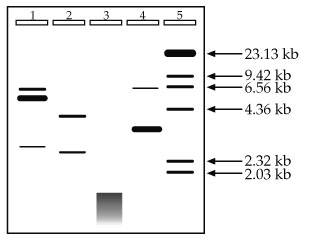
Agarose gel electrophoresis of labeled DNA in which the same ...
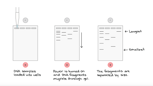
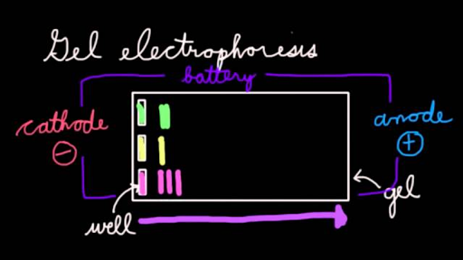
Gel electrophoresis (article) | Khan Academy

Reducing SDS-PAGE gel electrophoresis showing the light and ...

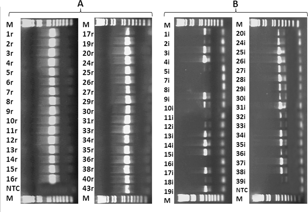
Mutagenesis efficiencies determined by restriction assays. A ...
Gel electrophoresis

Electrophoresis on agarose gel and polymerase chain reaction ...

InDesign Labeling / Annotating PCR Gel Pictures - Advanced Tutorial (Part 12)
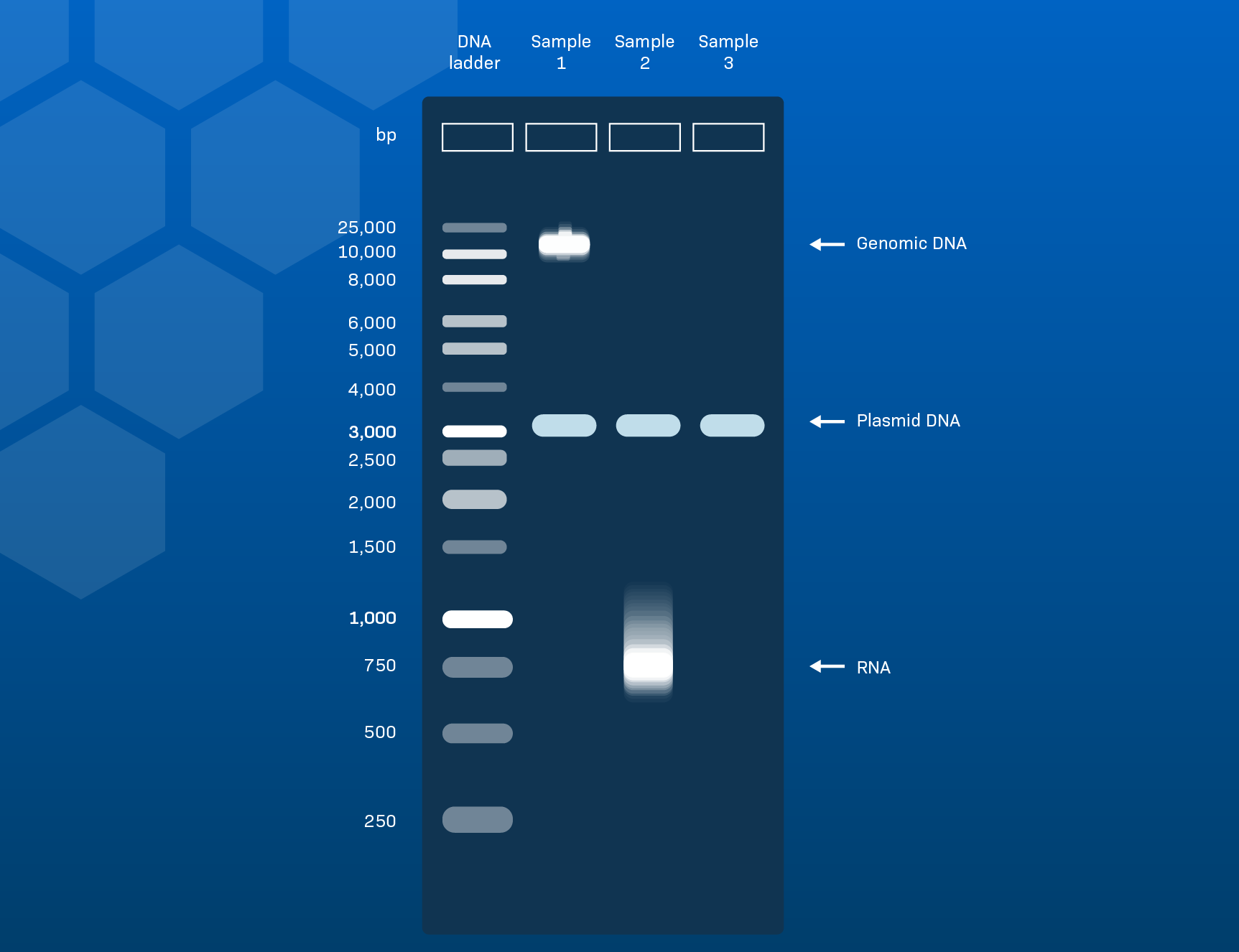
How to Interpret Agarose Gel Data: The basics - LabXchange

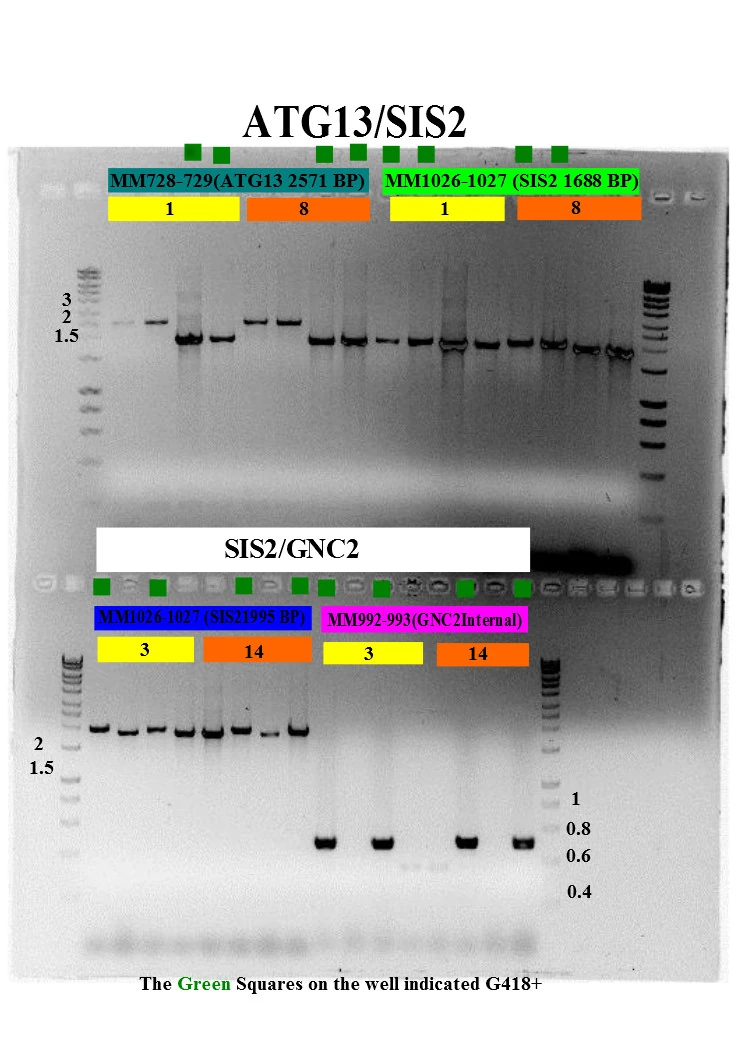
PCR amplification for single-site mutagenesis. A) Agarose gel ...

Synthesis of site-specific spin-labeled RNA. (A) Native gel ...

How to Read, Interpret and Analyze Gel Electrophoresis Results?

Invitrogen™ E-Gel™ EX Agarose Gels, 1%

ImageJ for Editing & Labelling PCR Gel Image | Biotechnology ...

CHAPTER 10 Flashcards | Quizlet
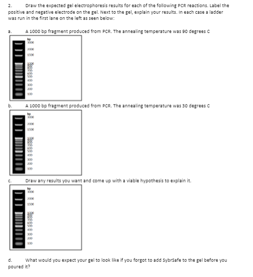
2. Draw the expected gel electrophoresis results for | Chegg.com
Gel Electrophoresis Assignment 1

ImageJ for Editing & Labelling PCR Gel Image | Biotechnology

Based on the data they collected using gel electrophoresis ...
Tutorial ImageJ
Agarose gel electrophoresis (AGE) image of the PCR products ...
A guide to lab reports
Texture analysis in gel electrophoresis images using an ...
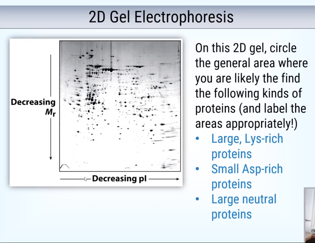
Solved on this 2d gel, circle the general area where you are ...

Label-free LC-MS and 2D gel electrophoresis, complementary ...
Electrophoresis
Molecular Vision: Wang, Mol Vis 2012; 18:3049-3056. Figure 2

DNA Sequencing
Chapter 2 Lab Overview and Background Information – BBS OER ...
Agarose gel electrophoresis - Wikipedia

Label-free Kinase Profiling Using Phosphate Affinity ...

InDesign Labeling / Annotating PCR Gel Pictures - Advanced ...
Annotating A Gel | Get Your Science On Wiki | Fandom
Solved] Lab 9 - Agarose Gel Electrophoresis 18.On the diagram ...

Agarose gel electrophoresis of RT- PCR products. The figure ...

Given below is the apparatus of agarose gel electrophoresis ...

Agarose Gel Electrophoresis: Results Analysis Video

Difference Gel Electrophoresis - an overview | ScienceDirect ...

Annotating Gels, Aligning text, and saving to a file
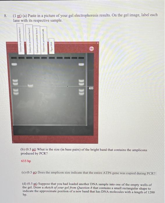
8. (1 B (a) Paste in a picture of your gel | Chegg.com
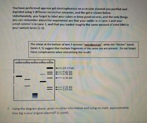
Solved You have performed agarose gel electrophoresis on a ...

Quantitative Protein Profiling Using Two-dimensional Gel ...

Agarose gel electrophoresis of labeled DNA in which the same ...
A guide to lab reports

2-D Difference Gel Electrophoresis – an accurate quantitative ...
Post a Comment for "44 how to label gel electrophoresis images"